By: medekomadmin
How 3D Anatomy Education Is Transforming Medical Learning Through Virtual Dissection
September 28, 2026
Anatomy is a three-dimensional subject. The human body is made with the arrangement of muscles, nerves vessels, organs and tissues that exist in a composite dimension with one another. Most anatomy education traditionally begins with two-dimensional diagrams, textbooks, figures and fixed images. For medical students, it is insufficient to simply memorize the names of these structures; they need to understand their location, how they relate to each other and their morphology when seen from different angles. It is at this point that 3D anatomy education and virtual dissection are opening up new opportunities for medical learning.
Interactive digital anatomy programs enable learners to examine anatomical structures from various angles, to go through the different layers, to look at cross-sections and to link their theoretical knowledge of anatomy with medical imaging. For example, institutions such as the University of Connecticut use virtual anatomy technology in conjunction with cadaveric dissection in order to help students make the connection between gross anatomy and cross-sectional anatomy as well as radiologic imaging and pathology.
In the context of medical education this link between anatomy and imaging is especially relevant.
New methods for exploring three-dimensional structures include 3D anatomy, virtual dissection, sectional imaging, and clinical cases. For them, this opens up more possibilities of visualizing, going over and examining complex anatomy. For teachers it can serve as a further tool for use in demonstrations, classroom discussions and for self-directed learning. For medical institutions it also provides the opportunity of setting up more flexible and interactive anatomy learning environments.
As medical education keeps on developing, the possibility of studying the human body in a digital way may turn out to be an ever more useful element in how anatomy is taught and understood.
What makes anatomy hard to visualize?
A major challenge in the study of anatomy is forming an accurate mental image of the human body. A textbook may name a nerve, blood vessel, or muscle, but it is only through spatial thinking that one can understand its topographical relationship and thus, form its clinical correlation. For example, merely learning the name of a structure is only one aspect of the process; a medical student might also be required to:- Where the structure is located
- What lies above or below it
- Which structures surround it
- How it appears in cross-section
- How it relates to nearby vessels and nerves
- How the anatomy connects with clinical imaging
From Static Anatomy to Interactive 3D Anatomy
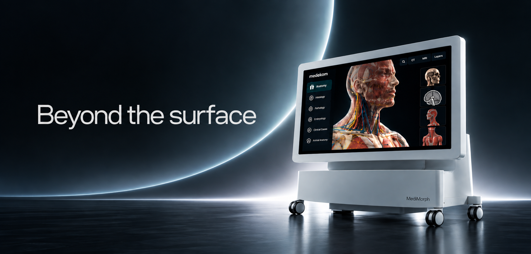
The technology for 3D anatomy alters the way in which learners interact with anatomical information. Rather than relying on a single static illustration, students have the option of using a digital anatomical model by rotating it, zooming in on areas of interest, progressing through the different layers and looking at the structures from various angles. The interactive method forms the basis of Medimorph's Virtual Anatomy Teaching Tool. The published material states that the programme combines 3D reconstruction with touch-based interaction and enables users to explore the human body, section by section and from various angles, in high definition. The touch control enables multi-angle slicing, rotation, zooming, and layer-by-layer virtual dissection. This is important because the study of anatomy cannot be understood from a single perspective. A student who is studying a particular anatomical area can look at the same structures from various angles and then go on to examine deeper layers.What Is Virtual Dissection?
Virtual dissection involves the use of interactive anatomical models and medical imaging as a means of studying anatomy. A virtual environment differs from a static diagram in that it enables learners to move through the various anatomical layers and to examine the structures over and over again. Research into the use of virtual dissection as an educational tool has been carried out together with traditional methods of teaching anatomy. What is important is that virtual dissection should not be simply seen as a replacement for all traditional methods of learning. Rather, it can offer an additional level of visualization and exploration. What this means for students is that they are able to go over troublesome structures, look at cross-sections and investigate anatomical relationships in a controlled digital environment, thus eliminating the need to imagine spatial placement and clinical correlation of complex anatomical structures.How 3D Anatomy Can Support Better Visualization
- The value of 3D anatomy is in the downstream activities that students can perform with improved understanding. Medimorph makes use of actual tissue-based data from cadavers and has see-through features which show the structures from more superficial layers down to the deeper tissues. It will enable students to go beyond just recognising a structure and instead look at how the various anatomical layers are situated in relation to one another.
- Learn anatomy from a number of different perspectives - Learners can view a structure from various angles by using rotation, zoom and multi-angle slicing. It is especially useful in cases where the spatial relationship of a structure is hard to understand from a single image.
- Perform virtual dissections - The gross human anatomy module offered by Medimorph allows for virtual dissections and cross-sectional studies. Learners can use repeated digital exploration as a means of going over anatomy whenever they are studying on their own or in the classroom.
- Link the anatomy with medical imaging - Anatomy studies take on greater clinical relevance when students are able to associate anatomical structures with diagnostic imaging. Medimorph contains over 1,700 CT/MRI scans and more than 180 real-world clinical cases. The clinical case materials consist of patient information, symptoms, diagnostic imaging and conclusions, as well as tools for exploring CT/MRI data and for carrying out 3D volumetric reconstruction.
"Where is this structure?"
to:
“What does the structure look like in a clinical image?”
More Than 3D Models: Building a Broader Anatomy Learning Environment
is not necessary for a powerful digital anatomy platform to limit itself to 3D visualization. The technical literature offered by Medimorph consist of over 3,000 three-dimensional anatomical structures, more than 3,000 sectional images, more than 100 educational videos, over 1,800 interactive exercises and more than 130 self-paced courses. The slice library also contains digital histology and pathology resources, allowing for adjustable magnification, annotations, and the designation of regions of interest. This creates a broader learning ecosystem in which anatomy can be explored through:- 3D structures
- Cross-sectional anatomy
- Histology
- Pathology
- CT/MRI imaging
- Clinical cases
- Videos
- Interactive exercises
- Self-paced learning
What can medical educators do with 3D anatomy technology?
Interactive anatomy can also be used to aid teaching besides individual study by students. Medimorph is intended for use in classroom demonstrations and is capable of connecting to projectors, smart TVs and other external displays when teaching in groups. The system is plug-and-play, mobile, and adaptable to various teaching workflows. It enables educators to introduce an anatomical structure into a classroom conversation and examine it with their students instead of depending just on a static image. For instance, a lecturer could show a specific anatomical area, rotate the model, go through the different layers, add annotations and then link the structure to sectional or clinical imaging. This results in a more interactive teaching experience while still keeping the educator at the heart of the learning process.What is Medimorph's place in modern medical education?
The platform offered by Medimorph combines a number of these digital learning features into one system. The Virtual Anatomy Teaching Tool features high-definition 3D anatomical visualization, interactive touch operation, virtual dissection, sectional studies and a wider medical resource database. The platform also includes aspects beyond gross anatomy such as embryology, animal anatomy, clinical cases and digital pathology. What this means for medical institutions is that digital anatomy can be incorporated into the overall teaching process rather than being confined to a single separate application.The Future of Anatomy Education Is More Interactive
Digital visualization, medical imaging, and interactive learning technologies are becoming more and more a part of medical education. It by no means has to involve giving up traditional teaching. The more useful question is:“In what way can digital tools aid the methods that medical students currently use to learn?”
Explore Interactive Anatomy Learning With Medimorph
Do you want to add interactive 3D anatomy, virtual dissection, and clinical imaging to medical education? Take a look at Medimorph and find out how digital anatomy technology can be used to support medical teaching and learning.
Explore Medimorph
Written by Yash Shah
Intern