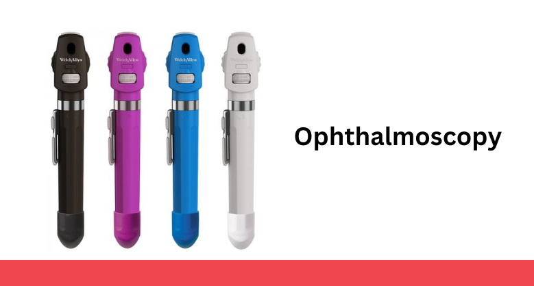
By: medekomadmin

Ophthalmoscopy

January 6, 2023

  • Ophthalmoscopy is a test to examine the back part of the eye called as fundus which is the light-sensitive area. The retina receives visual images and sends them to our brain's visual cortex through the optic nerve. The fundus consists retina, macula, optic disc, and blood vessels. Ophthalmoscopy also called fundoscopy is a part of an eye examination and is usually done during a routine eye check-up to screen for any eye diseases.
  • Types of ophthalmoscopy
  • There are three types of ophthalmoscopy and the test is performed accordingly- Direct ophthalmoscopy- This test is performed in a dark room using an instrument called an ophthalmoscope which has a small flashlight with several lenses showing a virtual, erect and 15 times magnified image.
  • Indirect ophthalmoscopy
  • Light attached in the head of the health care provider which allows the provider a wider view of the fundus of the eye. This test is usually done for viewing a detached retina. It produces real, inverted and 5 times magnified images.
  • Slit-lamp ophthalmoscopy- This gives the same view as indirect ophthalmoscopy but with higher magnification. An instrument called a slit-lamp is placed in front where the chin and forehead are put to make a steady position. Microscope part of slit-lamp is placed in front of the eye to see the view just like indirect ophthalmoscopy but with greater magnification.
  • Where this test is used?
  • Generally, it is a part of the routine test or in a complete eye examination which is mainly done by an ophthalmologist. But also detect-
  • Eye diseases like Glaucoma
  • Rental Detachment
  • Any damage to the optic nerve
  • Cytomegalovirus (CMV) retinitis (infection on the retina)
  • Melanoma is a type of skin cancer which can reach the eye
  • Patients showing signs and symptoms of hypertension, diabetes and any other disease related to blood vessels.
  • Preparation before ophthalmoscopy
      Eyedrops before performing indirect ophthalmoscopy for dilation of a pupil which may be slightly uncomfortable but are not painful. Direct ophthalmoscopy and slit-lamp ophthalmoscopy do not require eye drops. Dilation of the pupil makes eyes larger and easier for a check-up. Before using eye drops make sure to talk about your allergic problems or your ongoing medicines. The person with glaucoma or having a family history of glaucoma must tell the doctor about it.
  • Risk Multiple images are seen after turning off the light.
  • Eye drops may cause dry mouth, vomiting, nausea, flushing etc.
  • Precaution
  • Wear sunglasses after using eye drop to protect your eye.
  • Do not drive after the test are performed.
  • -Muskan Jain