By: medekomadmin
PNEOUMOTHORAX
September 20, 2023

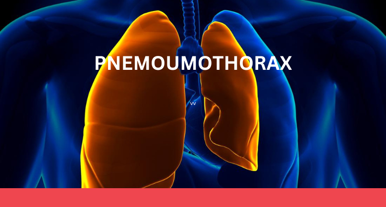
Pneumothorax is an accumulation of air into the pleural cavity i.e., between the lung and chest wall. This accumulation of air applies pressure which makes it collapsed lung. The lung can collapse completely or only a part of it can collapse which shows symptoms like shortness of breath, and pain in either of the chest.
Causes and types of pneumothoraxes-
- Depending on the cause or effect, pneumothoraxes can be categorised in several ways:-
- Rib fracture or broken ribs
- Diving, flying or at high altitude.
- Bullet wound
- Injury in the chest from sports or vehicle accidents.
- Smoking
- Pregnancy
- Marfan syndrome (connective tissue disorder)
- Family history
- A healthy person with a tall, thin body
- COPD (chronic obstructive pulmonary disease)
- Asthma
- Cystic Fibrosis
- Tuberculosis
- Severe ARDS (acute respiratory distress syndrome)
- Collagen vascular disease
- Pneumonia
-
- Simple- In such a case, the structures will remain intact .
- Tension-The position of other structures, like heart is generally affected. It tends to occur in people with chest pain, changes in pressure, any penetrating injury etc.
- Open-It happens in an open wound where air moves in and out of the chest.
-
- Sharp, sudden chest pain
- Rapid beathing or shortness of air
- Rapid heart rate
- Low blood pressure
- Respiratory discomfort
- Cardiac arrest
- Pathophysiology
-
-
- The pressure gradient inside the thorax cavity contains the lungs, heart, and blood vessels. Serous fluid is present between the parietal and visceral membranes which protect our lungs from friction. Normally lungs are inflated because the pressure inside them is negative as compared to atmospheric pressure. Any injury in the chest will cause air into the pleural cavity which increases the pressure equal to the pressure outside which causes the lungs to deflate.
- Symptoms of pneumothorax can be inconclusive in different types of pneumothoraxes. Generally, chest X-ray, computed tomography (CT), and ultrasound are used for diagnosing in which chest x-ray is most common. Recent diagnostic tools for pneumothorax include extended focused abdominal sonography for trauma (E-FAST).
- Complicity varies among individuals depending on the severity and treatment of people. Some complications are life-threatening and may lead to death. Some complications are- respiratory failure, pulmonary edema, heart attack, inability to breathe etc.
-
-
-
- The aim is to release pressure on the lungs and allows them to re-expand. The treatment of pneumothorax depends on the severity of the symptoms and the patient’s condition.
- Needle aspiration- A hollow needle with a small tube is inserted between the ribs into the pleural space and air should be aspired with the help of the needle.
- Chest tube - Chest tube may also be used to remove the air from the pleural cavity.
-